![]()
![]()
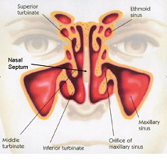
Septoplasty is a procedure designed to correct the shape of the nasal septum. To understand the goal of the procedure, you must first gain an understanding of the anatomy of the nose, nasal cavity, and nasal septum.
The nasal cavity is the space behind the nostrils extending back to the upper throat. It opens into the area of the throat called the nasopharynx and is divided into right and left sides by the nasal septum. Abnormalities of the nasal septum are a major cause of blocked nasal airway and nasal congestion.
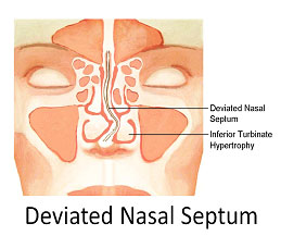
As you can see above, nasal septal deviation can block the nasal airway making air movement through the nasal cavity difficult or impossible. These deformities can be present in minor form since birth and increase in severity as the facial bones mature, or they can be caused by sudden trauma which will fracture the nasal septum. Usually, a person with a significant septal deviation will have congestion worse on one side of the nose. However, septal deviation isn’t always simply to the right or left. The septum can be bent in several locations causing blockage of both sides of the nasal cavity.
Whatever the cause, if the deviation is bad enough to cause discomfort from blocked nasal airway, surgery can be considered.
Septoplasty is the name of the operation used to correct deviations of the nasal septum. Like any other type of plastic surgery, care must be taken to remove as little native tissue as possible while achieving the appropriate change in the shape of the abnormal structure. In case of the nasal septum, the goal is to save the pink lining covering the septum and concentrate all efforts on correcting the deviations of the septal skeleton.
As you can see above, the skeleton of the nasal septum is made of both bone and cartilage. The cartilage is in front (blue
 in the diagram above) while the bony septum is in back. Deviations of the cartilaginous require more attention to detail and meticulous surgical technique because the cartilaginous septum supports the soft part of the lower nose (just above the tip of the nose). If too much cartilage is removed or damaged in this area, the area above the nasal tip will collapse and you end up with what is called a “saddle nose” deformity. The most severe saddle nose deformities are caused by large septal perforations. In these cases, a large hole forms in the cartilaginous portion of the nasal septum as the cartilage and the covering lining are lost. Below is an example of a saddle nose deformity.
As you can see to the left, the skeleton of the nasal septum is made of both bone and cartilage. The cartilage is in front (blue in the diagram above) while the bony septum is in back. Deviations of the cartilaginous require more attention to detail and meticulous surgical technique because the cartilaginous septum supports the soft part of the lower nose (just above the tip of the nose). If too much cartilage is removed or damaged in this area, the area above the nasal tip will collapse and you end up with what is called a “saddle nose” deformity. The most severe saddle nose deformities are caused by large septal perforations. In these cases, a large hole forms in the cartilaginous portion of the nasal septum as the cartilage and the covering lining are lost. To your right is an example of a saddle nose deformity. The basic technique of the operation is sequenced as follows: First, the lining over the bony and cartilaginous septum is elevated on both sides. Next, various minimally invasive techniques are used to reshape, sculpt, and realign the deviated portions of the nasal septum. In areas that can tolerate los of tissue, severe deflections of bone can be removed. Once the septal skeleton is in the appropriate middle position, the lining flaps are returned to their normal position and sutured in place. In almost all cases the patient is discharged home the same day.
|
Dr. K. Amini 8435 Reseda Blvd Northridge, CA 91324 |


@ ENT Doctor LA, Website was created and maintained by Biago Media