![]()
![]()
 Swelling of the lining around the sinus can be caused by infection, allergies, or anatomical blockage. This can cause the sinus outflow tract to close and prevent the sinus from emptying itself of its mucous. That mucous will fill the sinus and become a site for bacteria to grow, eventually leading to a sinus infection. The immune response may lead to more swelling, resulting in more backup of mucous and a chronic infection that keeps cycling over and over.
Successful treatment of chronic sinusitis involves breaking the cycle of swelling and infection. To do that, you have to treat whatever factor caused the initial swelling to begin with, and also treat the active infection that helps keep the cycle going. When medical management is not successful in breaking the cycle, endoscopic sinus surgery is offered.
To achieve the goal of cure surgically, the sinus drainage pathway must be opened to allow the sinus to drain. This seemingly simple goal must be reached carefully in order to keep the sinus functioning properly once it is surgicaly opened. To understand this last concept, you have to remember that the sinus lining functions to produce mucous and then move it out in an orderly fashion. The need to preserve that mucosa and its ability to expel the excess fluid and mucous out of the natural sinus outflow tract is one of the main goals of functional endoscopic sinus surgery. The image to the left is that of a microdebriding instrument. It allows for a controlled, careful removal of the minimal amoiunt of bone and mucosa that is necessary in order to reestablish a normal sinus drainage pathway. It is
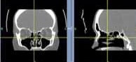
guided into the nasal cavity with the help of a nasal endoscope which provides a magnified view of the intranasal and sinus anatomy. To the right, you see a typical image of a patient's CT scan in coronal and in sagital views when utilizing intraoperative Image Guided Navigation System. These systems allow real time localization of the instruments that are used during sinus surgery. The cross hatch on the image could represent the tip of the microdebrider shown to the left. That way, you know where the instrument is with much more certainty. This helps to limit complications.
|
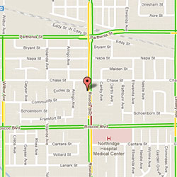
Dr. K. Amini 8435 Reseda Blvd Northridge, CA 91324 |


@ ENT Doctor LA, Website was created and maintained by Biago Media